+1.310.385.1918 | 8436 W. 3rd St. #800 L.A.
Follow us:
![]()
![]()
![]()
![]()
![]()
+1.310.385.1918 | 8436 W. 3rd St. #800 L.A.
Follow us:
![]()
![]()
![]()
![]()
![]()

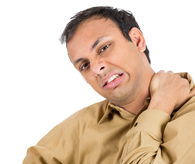
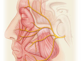
Hemifacial spasm (HFS) is a disorder characterized by uncontrollable twitching or spasms of one side of the face. Its onset is spontaneous, most commonly consisting of subtle twitching of the muscles around the eye (orbicularis oculi). Over time, this can progress to involve the other muscles innervated by the facial nerve. In the extreme situations HFS can be disfiguring and interfere with a person’s lifestyle. At times the spasms can be quite painful. The cosmetic issues and the understandable psychological results can result in social disability or isolation. As the disease progresses there can be weakest of the muscles of the face, which can be irreversible. In most cases the spasms are increased with anxiety and activity.
While there are different diseases that can be related to twitching of the face, HFS refers to spasms that are spontaneous in onset and not following an identifiable disorder such as Bell’s palsy or a viral infection. It is felt that most of these cases are caused by compression and injury of the facial nerve where it exits the brainstem by an artery (vascular compression). It is theorized that the pulsations of the artery leads, in time, to erosion of the insulation of the nerve (the myelin covering) and a “short circuiting” of the electrical impulses (ephaptic transmission).
When minor twitching of the eye is seen, no treatment may be required. However, as the disease progresses most patients seek medical intervention. In many cases Botulinum Toxin (Botox), injections into the effected facial muscles can result in good control of the spasms. However, this is not permanent and requires repeat injections after several months.
In many patients permanent treatment of HFS can be achieved with surgery. The preferred surgical treatment is known as a microvascular decompression (MVD) procedure. A small incision is made behind the ear and a nickel sized opening in the skull allows visualization of the area of the facial nerve and brainstem, the cerebello-pontine angle (CPA). Using a high powered microscope and endoscopes the blood vessel compressing the facial nerve is lifted and a small pad or sponge is placed between it and the nerve, acting as a shock absorber. The pulsations of the artery are no longer eroding the covering of the nerve. Excellent relief of facial spasms is achieved in more than 90% of properly selected patients. Hospitalization is typically 2 days.