+1.310.385.1918 | 8436 W. 3rd St. #800 L.A.
Follow us:
![]()
![]()
![]()
![]()
![]()
+1.310.385.1918 | 8436 W. 3rd St. #800 L.A.
Follow us:
![]()
![]()
![]()
![]()
![]()

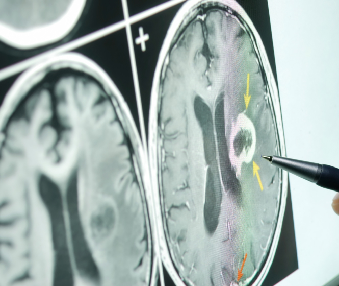
Technological advances during the last 20 years have allowed less invasive procedures in most of the surgical specialties. Smaller incisions and less operative trauma and manipulations mean shorter surgery, reduced hospitalization and faster recuperation following surgery. Recently, these techniques have been applied to diseases of the brain. Using endoscopes and high powered microscopes, there has been tremendous improvement in our ability to treat lesions such as pituitary tumors, trigeminal neuralgia, tumors of the skull base and problems with the ventricles of brain (eg colloid cysts).
Small telescopes (endoscopes) to treat various disorders have been utilized for much of the last 4 decades. However, only recently have they been used in Neurosurgery. Endoscopes are narrow tubes measuring 2.5-4.5 millimeters that consist of a series of glass lenses or fiber optic strands. Because of the delicate nature of the central and peripheral nervous systems these endoscopes are significantly smaller than those used in other surgical specialties that more commonly measure 5-10 millimeters in diameter. The scope is attached to a camera, allowing the surgeon to visualize the image in front of the endoscope on a high resolution television screen. Some endoscopes are flexible or steerable making it possible to navigate into areas that would be otherwise difficult to access using traditional means. Endoscopes can be used alone (endoscopic or endoscope controlled surgery) or in conjunction with the microscope (endoscope-assisted surgery). Typically endoscopic techniques allow much smaller incisions to gain access to the brain, spine and peripheral nerves (eg carpal tunnel syndrome).
Many lesions involving the pituitary gland and skull base can be excised through the nose endoscopically. By exposing the back of the nose, through one or both nostrils, and the adjacent paranasal sinuses (transsphenoidal sinus) the region of the pituitary gland and cavernous sinus can be visualized. Most commonly this approach is used to treat neuroendocrine tumors (pituitary adenomas, rathke’s cysts and craniopharyngiomas). However, selected skull base tumors such as meningiomas, chordomas and various invasive cancers can be successfully removed through this route (extended transsphenoidal approach).
Many tumors that previously required larger procedures can be removed through a small incision in the eyebrow. The operative microscope and the rigid endoscope are both utilized. Most commonly skull base tumors, such as meningiomas and craniopharyngiomas, located underneath the frontal lobe of the brain are good candidates for this type of surgery. The advantage is that eyebrow surgery requires less manipulation of the brian and reduced inherent injury to the brain, skull, and scalp. The cosmetic results are also quite acceptable when compared to more invasive traditional surgery.